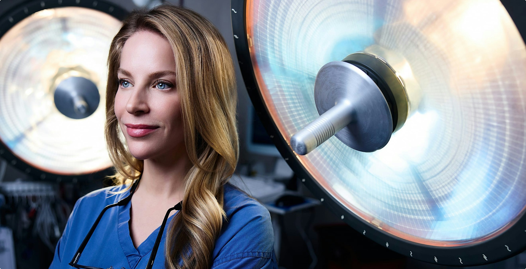
Precision-Driven Innovation for the Body’s Most Intricate Anatomy

Some brain and skull base conditions can now be treated using advanced techniques that avoid traditional craniotomies. Through the orbit, the natural corridor around the eye, Dr. Allison Coombs collaborates with neurosurgeons to provide access to targeted intracranial areas using minimally invasive, endoscopic-assisted approaches.

As one of the few oculofacial surgeons offering this technique, Dr. Coombs combines microsurgical skill, deep anatomical knowledge, and a collaborative approach to make high-stakes surgery safer and less invasive for select patients.

What Are Minimally Invasive Neurosurgical Approaches via the Orbit?

These techniques use the orbit (eye socket) as an access pathway to the skull base, cavernous sinus, or brain, avoiding large scalp incisions and brain retraction. Using an endoscope and microsurgical tools, Dr. Coombs helps create a precise, internal route that allows the neurosurgeon to reach and treat the target area. Conditions that may be approached this way include:

  • Skull base tumors
  • Optic nerve sheath meningiomas
  • Cavernous hemangiomas
  • Vascular lesions or aneurysms
  • Inflammatory or infectious masses
Banner media
woman facing the right with her eyes closed and hair in a bun

How Does This Approach Work?

The procedure begins with a detailed plan using CT and MRI imaging. Dr. Coombs carefully dissects the orbital tissues through a hidden eyelid incision or conjunctiva. Working alongside a neurosurgeon, she provides access through the orbital apex or medial wall.

Once the neurosurgical portion is complete, Dr. Coombs ensures closure of the orbital tissues and meticulous wound care to protect the eye and restore anatomy.

What Are the Benefits of This Minimally Invasive Approach?

By avoiding traditional craniotomy, these techniques reduce trauma to the brain and surrounding tissues. Additional benefits of this innovative approach include the following:

  • No large scalp or forehead incisions
  • Less disruption to brain tissue
  • Faster recovery and reduced hospital stay
  • Lower risk of complications in select cases
  • Preservation of facial aesthetics
  • Enhanced visualization through high-definition endoscopy

Who Is a Candidate for This Approach?

Not all intracranial conditions can be treated this way, but for select lesions near the orbit or skull base, this route offers major advantages. The following are ideal candidates:

  • Patients with orbital apex or anterior skull base lesions.
  • Those who require a biopsy or targeted mass removal.
  • Individuals with stable systemic health and anesthesia clearance.
  • Patients who are seeking less invasive surgical alternatives.
  • Candidates referred by a neurosurgeon for collaborative care.
Background media
Doctor media

Dr. Allison V. Coombs


Why Patients and Physicians Go to Dr. Allison Coombs

Few surgeons are trained to navigate both the orbit and skull base with the delicacy these procedures demand. Dr. Allison Coombs is board-certified and fellowship-trained in oculofacial plastic surgery and ophthalmology, giving her unmatched expertise in this highly specialized area.

She completed her advanced training at Tufts, Weill Cornell, and MEETH, where she now leads the oculofacial plastic surgery division. Dr. Coombs collaborates with neurosurgical and ENT specialists to deliver seamless, interdisciplinary care in some of the most complex cases. Her patients and colleagues go to her for technical excellence, calm confidence, and a deeply human approach to care.

Your Consultation

If you’ve been referred for a minimally invasive skull base or orbital procedure, your consultation with Dr. Coombs will include a detailed review of your imaging, discussion of goals, and a clear explanation of the collaborative surgical plan. She will walk you through what to expect before, during, and after the procedure, and coordinate with your broader medical team as needed.

Concerning Costs

Because these procedures are often medically necessary and highly specialized, many are covered by insurance. Costs vary depending on the complexity of the procedure, the surgical facility, and coordination with other surgical specialists. Our team will assist with benefit verification and cost transparency at your consultation.

Minimally Invasive Neurosurgical Treatment Procedure

This surgery is typically performed in a hospital setting under general anesthesia. It may involve multiple specialists depending on the area in need of access. The following is what to expect during the procedure:

  • Preoperative imaging, surgical planning, and team coordination.
  • Access created through the eyelid or conjunctiva by Dr. Coombs.
  • Endoscopic tools are used to reach the skull base or target area.
  • Neurosurgical or ENT procedure performed through the access corridor.
  • Closure and reconstruction of the orbital tissues by Dr. Coombs.
  • Postoperative monitoring and follow-up with all involved specialists.

Schedule A Consultation

Contact Us
Contact us media

Recovery and Results

Recovery varies based on the condition treated, but most patients experience less postoperative pain and faster healing than traditional craniotomies. Dr. Coombs monitors orbital healing closely, while your neurosurgeon oversees intracranial recovery. Swelling and bruising around the eye typically resolve within one to two weeks.

Banner media

Types of Minimally Invasive Neurosurgical Techniques via the Orbit

  • Transorbital Endoscopic Skull Base Surgery
  • Optic Nerve Sheath Decompression or Biopsy
  • Orbital Apex Mass Removal
  • Combined ENT/Neurosurgical Tumor Resection
  • Biopsy of Intraconal or Retrobulbar Masses

Related Procedures

  • Traditional Craniotomy – May be required if the lesion is not accessible via the orbit.
  • Orbital Tumor Removal – For primary orbital lesions.
  • Orbital Decompression – If space needs to be created for access.
  • Endonasal Endoscopic Surgery – For sinus or pituitary tumors.

FAQ Minimally Invasive Neurosurgical Approaches

Is this safer than traditional brain surgery?

For certain conditions, yes. It avoids large openings and reduces brain retraction, lowering certain risks.

Will this affect my vision?

The goal is to preserve or improve vision. Dr. Coombs prioritizes the protection of optic structures during access.

Do I need to see a neurosurgeon first?

Often, yes. Dr. Coombs works in close collaboration with neurosurgeons to determine if this approach is appropriate.

Is recovery faster than a craniotomy?

Typically, yes. Patients often return to activity sooner with less postoperative discomfort.

Contact us media
Accessibility: If you are vision-impaired or have some other impairment covered by the Americans with Disabilities Act or a similar law, and you wish to discuss potential accommodations related to using this website, please contact our Accessibility Manager at (212) 702-7733.
Contact Us